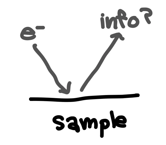
After finishing electrochemically testing a cell, I have the option to disassemble it and view components in a scanning electron microscope (SEM). Unlike an optical microscope that uses visual light, SEM uses an electron beam. Using electrons allows us to see at smaller scales, due to electrons having a smaller wavelength than visible light. An image is created based on how electrons interact with and are reflected by the sample. In an anode sample, I typically look for any cracks or gaps in the anode surface. A smoother anode surface typically signifies more uniform ion deposition and better interface contact.
In conjunction with SEM, I also use energy x-ray spectroscopy (EDS) to identify elements present on the sample surface. This can give us insight into chemical reactions taking place. For example, we might discover that we likely have two elements forming a chemical compound based on the ratio of their elements. Or we might be able to figure out where the original solid electrolyte interface broke using our knowledge of the components of the anode soak solution. X-ray photoelectron spectroscopy (XPS) is actually better suited for this task of identifying elements present at the surface but has more restrictions on which solvents are safe for use, so I use both techniques.
Through this testing, we really want to learn more about the battery mechanisms. Since handmade coin cells are often not very reproducible or scalable, expanding the understanding of battery science is probably the most important role that academic battery researchers play. If we can understand battery science and the limitations better, we can ultimately develop better performing cells.
